Published in May 1988
TREATMENT OF PERIODONTAL LESIONS IN MULTIROOTED ELEMENTS 
GUSTAVO 
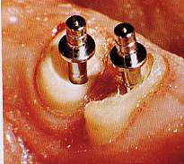
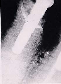
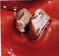
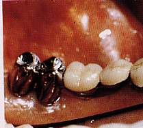
PETTI
Physician and Surgeon specializing in Dentistry. Periodontist.
Piazza Repubblica 4, 09129 Cagliari, Italy.
tel ++39 070 498159, fax ++39 070 400164
web site www.gustavopetti.it

Graft 
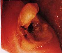
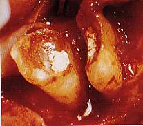
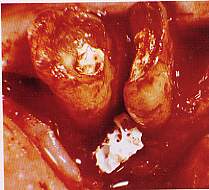
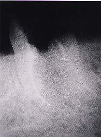
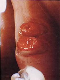
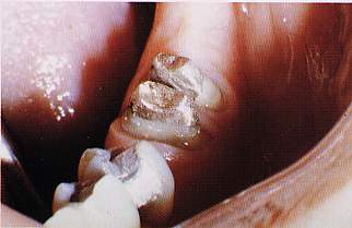
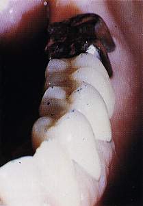
with heterologous bone

Figs 20, 21. 3rd degree lesion of the bifurcation of 4.7 to which is associated an endoperiodontal lesion.
Fig. 22. After detaching a mucoperiosteal flap the roots are separated.
Fig. 23. Following bone remodelling the operation continues with an interradicular graft of Kiel bone to correct the vast bone crater.
Figs 24, 25. Once tissues have healed, two teeth with provisory cores are constructed.
Figs 26, 27. Two cast gold cores are made. They act as the terminal pillar of a fixed gold-platinum-porcelain prosthesis restoring the lower right arch.
Fig. 28. X-ray four years after the operation.
Fig. 29. 3.7 with the floor of the pulp chamber completely destroyed by the process of dental decay.
Fig. 17. Positioning of shaped Interpore 200.
Fig. 30. After sculpting a mucoperiosteal flap the roots are separated.
Fig 32
Fig 31
Fig 33
Figs 31, 34. After healing, the two cores are constructed with Duralay and cast in gold respectively.
Fig 34
Fig. 35. Finally, the lower left side is restored with the prosthesis.