 |
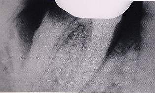
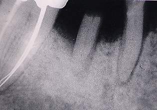
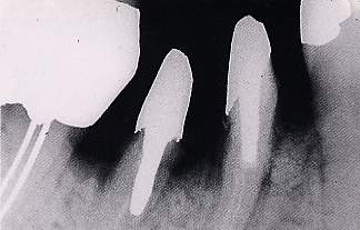
| Fig. 7. X-ray
of 4.6, which shows severe periodontal damage. |
|
|
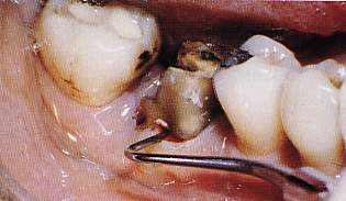 |
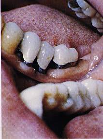
| Fig. 8. A passing
3rd degree lesion is probed. |
|
|
|
|
|
|
|
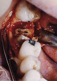 |
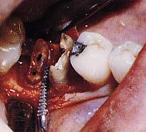
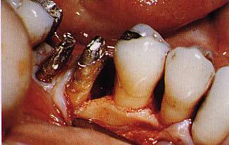
| Fig. 9. After
sculpting a mucoperiosteal flap, the roots are separated. |
|
|
|
|
|
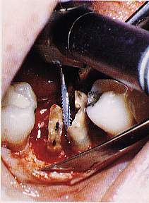 |
| Fig. 10. The bony
crest around the roots is lowered. |
|
|
|
|
|
|
|
|
|
|
|
|
 |
| Fig. 11. The interradicular
furrows are created with a file. |
|
|
 |
| Fig. 12. An X-ray
is taken to ensure that all protrusions have been eliminated. |
|
|
|
|
|
|
|
|
|
|
|
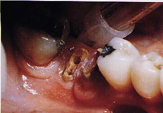 |
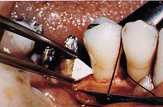
| Fig. 13. The
roots can now be worked on. |
|
|
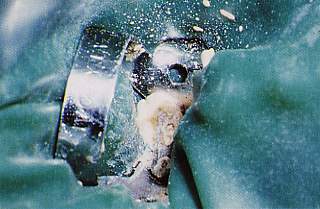 |
| Fig. 14. The dam
can be positioned and the endodontal treatment can proceed. |
|
|
|
|
|
|
|
|
|
|
|
 |
|
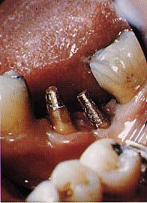
| Fig.
15. Prosthesically, the roots are restored with two cast gold
cores. |
|
|
|
 |
| Fig. 16. Then
the treatment of the mesial bone defect at 4.6 begins with a
graft of Interpore 200. |
|
|
|
|
|
|
|
|
|
|
|
 |
| Fig. 17. Positioning
of shaped Interpore 200. |
|
|
 |
| Fig. 18. X-ray
of the bone defect following reconstruction after the graft. |
|
|
|
|
|
|
|
|
|
|
|
 |
| Fig. 19. Definitive
gold-platinum-porcelain prosthesis. |
|
|
|
|
|
|
|
|
|
|
|
|
|
|