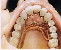
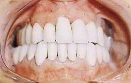
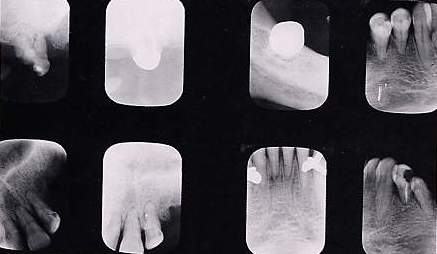
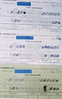
PERIODONTAL TREATMENT PRIOR TO PROSTHESIS
GUSTAVO PETTI
Physician and Surgeon specializing in Dentistry. Periodontist.
Piazza Repubblica 4, 09129 Cagliari, Italy.
tel ++39 070 498159, fax ++39 070 400164
web site www.gustavopetti.it
Fig. 17 Terminal gold-platinum-porcelain prosthesis Fig. 18 Enlargement of details of the intracoronal attachment for MacCollum-type horizontal retention ceramics in an intermediate position and actual milling with the drive bars to the left of the prosthesis.
Figs. 19-20 Extra-coronal "T" attachment with male body and screw to regulate friction (Fig. 49) and a reducible gold-platinum matrix for ceramics (Fig. 50) with milling and drive bars on the right side of the fixed and mobile prostheses.
Figs. 21-22 Upper and lower prostheses positioned in the mouth.
Fig. 23 Orthopantomography performed prior to periodontal treatment. Fig. 24 X-rays top left: 1.7 before and after implant; bottom, 2.1 and 2.2 before and after implant (second follow-up after 13 months). Top and bottom right: bone situation of teeth in lower arch with no important lesions.
Fig. 26 Same measurements (first and last) transferred onto graph paper
Fig. 25 Details of the three periodontal clinical records with measurements of pockets taken at the time of the first examination, periodontal re-evaluation following initial preparation (three months later) and at the end of treatment.