REALIGNMENT OF INCISORS
GUSTAVO PETTI
Physician and Surgeon specializing in Dentistry. Periodontist.
Piazza Repubblica 4, 09129 Cagliari, Italy.
tel ++39 070 498159, fax ++39 070 400164
web site www.gustavopetti.it

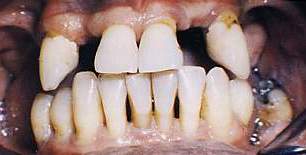
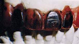
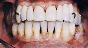
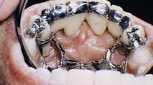
BONE REHABILITATION OF INCISORS

PERIODONTAL TREATMENT PRIOR TO PROSTHESIS

REALIGNMENT OF INCISORS

COMPLETE REHABILITATION OF HALF AN ARCH

OBJECTIVES

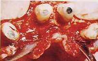
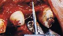
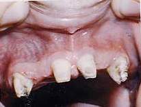
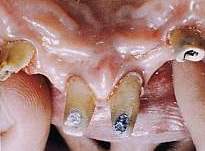
  • Reorganization of pillars for rehabilitation of incisors
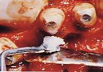
  • Reconstruction of gum adhering to 2.3
  • Follow-up to verify cure
Treatment of bone defects
Fig. 1 Initial situation.

OBJECTIVE
Adjust pillars for rehabilitation of incisors


Fig. 2 Study with models to obtain a realignment of incisors The wax model prepared by enlarging the central teeth and keeping only the lateral one as a bridge shows a poor result
Figg.3-4 It is thus decided to use the space destined for the lateral to insert, as a bridging element, the lateral and the canine, thus transforming the natural canine into the first premolar.
Fig. 5 Following curettage and scaling, the operation proceeds with osteotomy and osteoplastics with an osteotribe away from the roots and scalpels near them. Fig. 6 The bone defects palatally circumferential to the central teeth and a hemiseptum mesial to 1.3 are shown.
Figg. 7-8 Reconstruction work proceeds with a Periograft. The wound is sutured with single stitches and, once the prosthesis has been inserted, the area of the operation is protected.
Figg. 9-10 Vestibular and palatal healing after 18 months. Then the Duralay cores are created and cast in gold on the canines to better transform them into premolars (also considering the serious coronal damage present).

OBJECTIVE
Reconstruct a sufficient band of gum adhering to 2.3
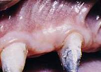
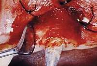
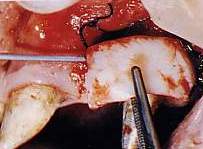
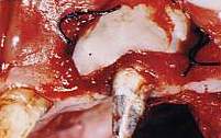
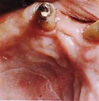
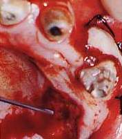
 

Treatment of gingival defects
Fig. 11 Insufficient adhering gum in the 2.3 area. Fig. 12 The receiving bed is prepared by suturing the previously detached mucous flap with safety stitches.
Figg. 13-14 Suturing of graft taken from the juxtagingival area of the palate with fibrin glue.
Fig. 16 Checking of donor site ninety days after operation.
Fig. 15 Protection of donor site with an amniotic membrane grafted with fibrin glue.
Fig. 
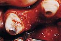
17 Checking of free gingival graft after ninety days.