|
PERIODONTAL
TREATMENT PRIOR TO PROSTHESIS
|
|
GUSTAVO PETTI
Physician and Surgeon specializing in Dentistry. Periodontist.
Piazza Repubblica 4, 09129 Cagliari, Italy.
tel ++39 070 498159, fax ++39 070 400164
web site www.gustavopetti.it
|
|
|
|
OBIETTIVI
- Treatment of bone defects of the upper arch. Construction of a fixed
prosthesis for the lower teeth and a prosthesis with attachments for
upper ones.
- To treat bone defects prior to application of a prosthesis.
|
|
|
|
|
|
|
|
|
|
|
|
|
|
|
|
|
|
|
|
|
|
OBJECTIVE
To treat bone defects prior to application of a prosthesis. |
|
|
|
|
|
|
|
|
|
|
|
|
|
|
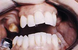 |
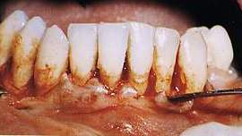
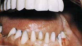
Fig.
1 Case at beginning. A chamfered mucoperiosteal flap will be sculpted on
the inside to eliminate the periodontal pocket and gain access to the bone
defects; then, following curettage and scaling in the mesial bone defect
at 1.7, osteoplastics will be performed with an osteotribe. |
|
|
|
|
|
|
|
|
|
|
|
|
|
|
|
|
|
|
|
|
 |
|
|
|
 |
 |
|
|
|
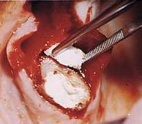
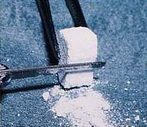
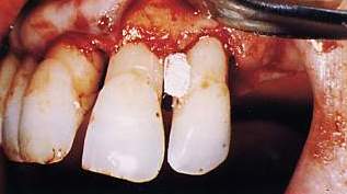
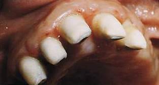
Fig.
3 A piece of Interpore 200 is prepared by modelling with a Beaver 64. |
|
Fig.
4 Interpore 200 implant in the bone defect. |
|
Fig.
2 Easily visible bone defect in three parts (which does not involve the
trifurcation) following curettage and bone remodelling. |
|
|
|
|
|
|
|
|
|
|
|
|
|
|
|
 |
 |
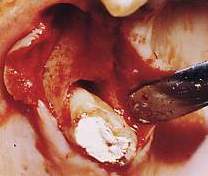
| Fig.
5 Bone implant seen mesially at 2.2 for the correction of a crater that
cannot be treated only with resective surgery and mesially at 2.1 for treatment
of a hemiseptum. |
|
|
|
|
|
|
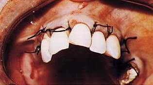
| Fig.
6 After modelling the implants on the palatal side as well, suturing is
performed with single stitches. After six months a cast gold core will be
created in 1.7. |
|
|
|
|
|
|
|
|
|
|
|
|
|
|
 |
|
|
|
|
|
|
 |
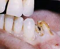
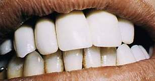
| Fig.
7 Avulsion is performed on 3.4 because of the severe periodontal damage. |
Fig.
8 Excision for the new attachment in the lower arch |
|
|
|
|
|
|
|
|
|
|
|
|
|
|
|
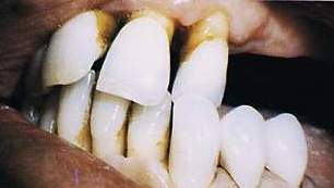 |
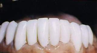
Fig.
9 A temporary prosthesis in 3.3-3.5 is created, maintaining crossbite |
|
|
|
|
|
|
|
|
|
|
|
|
|
|
|
|
|
|
|
|
|
|
|
|
|
 |
 |
| Figs.
10-11 Prosthesic preparation of upper teeth to realign them, correct crossbite
and stabilize them definitively with a temporary prosthesis with palladium
alloy bite to maintain the vertical dimension. |
|
|
|
|
|
|
|
|
|
|
|
|
|
|
 |
 |
 |
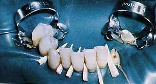
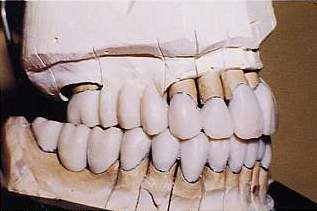
Figg.
12-13-14 The procedure is repeated for the lower arch |
|
|
|
|
|
|
|
OBJECTIVE
To construct a fixed prosthesis for the lower teeth and one with attachments
for the upper teeth
|
 |
 |
|
Fig.
15 A palladium alloy prosthesis, with rough modelling with resin as a test
piece, is constructed to check and record the necessary occlusal and gnathological
data. |
Fig.
16 The centricity, laterality and protrusive waxes are recorded |
|
|
|
|
|
|
|
|
|
|
|
|
|
|
|
 |
|
|
|
|
|
|
|
|
|
|
 |
|
|
|
|
|
|
|
|
|
|
|
|
|
|
 |
|
|
|
|
|
|
|
|
|
|
|
|
|