|
|||||||||||||||||
 |
 |
||||||||||||||||
|
|||||||||||||||||
|
|||||||||||||||||
 |
|||||||||||||||||
 |
|||||||||||||||||
| 3. The sutured free gingival graft; | 4. the graft covers the recession and increases the band of gum adhering around the canine. | ||||||||||||||||
| Periodontal
surgical methods
|
|||||||||||||||||
 |
 |
||||||||||||||||
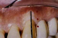
| 5. Flap with
coronal repositioning and free gingival graft: an incision is made along
the line of the mucogingival junction; |
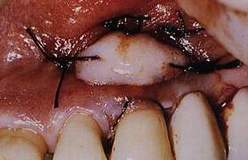
6. After freeing the tissue between the incision and the free edge, the flap is sutured with a suspended stitch in a more coronal position; | ||||||||||||||||
 |
 |
||||||||||||||||
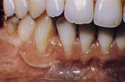
| 7. A free gingival graft (red in the drawing) is sutured. | 8. Gingival recession at 2.3 with a band of adhering gum. | ||||||||||||||||
 |
 |
||||||||||||||||
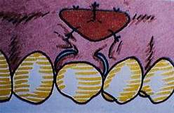
| 9. Upper left: the tissue between the free edge and the incision along the mucogingival line is freed. | 10. Upper right:
the flap is sutured coronally: thus a lozenge of tissue that will receive
the free graft is uncovered. |
||||||||||||||||
 |
 |
||||||||||||||||
| 11. Centre left: taking of the free graft. | 12. Above: graft sutured apically to the coronally repositioned flap. | ||||||||||||||||
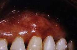 |
13. To the side: the recession has been covered and the adhering band of gum around it has increased. | ||||||||||||||||