 |
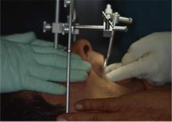
Fig. 22.
To ensure the success of oral rehabilitation, it is of fundamental importance to be able to transfer as accurately as possible the precision master patterns onto an articulator. The WhipMix facial transfer arch is quite useful and easy to use: the two posterior points of reference are obtained by inserting the terminals of the arch in the meatus acusticus externus; the third anterior point of reference is found by the glabellar support, which defines the anterior vertical position of the arch. In this way the hinge axis-orbital plane reference plane is defined. A horseshoe fourchet allows aligning of the upper and facial arches. In conclusion, having thus transferred the master patterns onto an articulator, we can orient the patterns of the arches with respect to the cranium and study the slope of the condylar paths and the inclination of the Bennett angle (Bauer Gutowski, 1984). |
||||||||||||
 |
|||||||||||||
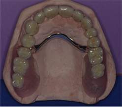
| Fig. 23. Combined gold-porcelain upper denture. Binat attachments with shock-absorbing hinges have been applied to the cores of 1.4 and 2.5. For the replacement of missing elements a palatal bar of the skeleton was designed to maintain the connection of the two cols on which the attachments and the missing elements of SR resin have been applied. |
|||||||||||||
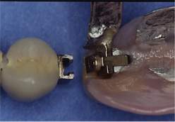 |
Fig. 24.
Enlargement showing the Binat attachments with a shock-absorbing hinge. |
||||||||||||
 |
 |
||||||||||||
|
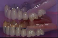
Figs 25, 26.
The hinge movement on the sagittal plane of the two posterior cols can be seen clearly. This movement, together with the shock-absorbing device, makes it possible to replace the missing back teeth with a prosthesis having a metal skeleton with precision attachments on implants, which would otherwise be impracticable. |
|||||||||||||
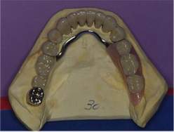 |
Fig. 27.
Combined gold-porcelain lower prosthesis on natural teeth and an ARS 88 prosthesis with a metal skeleton and precision attachments. From 3.5 to 3.8 a bar was designed to join 3.8 to the other elements since it was at risk and had been saved by elongating the clinical crown, since decay and fracture of the lingual wall were subgingival and below the bony crest, with an apically repositioned flap and periodontal osteotomy and osteoplastics, endodontal treatment and a gold core. If in future, but at the time of the latest checkup after 12 years it was found to be in perfect health, problems should arise, it would be sufficient to separate the bar between 3.5 and 3.8, which would be extracted. The advantage of this would be that the existing skeletoned prosthesis would require modification only by lowering it after healing. |
||||||||||||
|
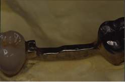
Fig. 28.
Detail of the bar with the attachment between 3.5 and 3.8. |
 |
||||||||||||
 |
 |
||||||||||||
|
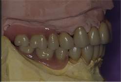
Figs 29, 30.
The prosthesis on the patterns seen frontally and laterally. Note the good overjet and overbite, which contribute both to functionality and appearance. |
|||||||||||||
 |
 |
||||||||||||
|
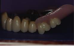
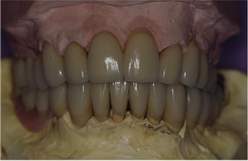
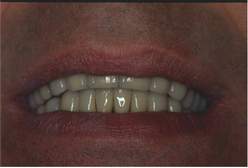
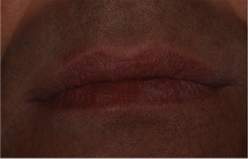
Figs 31, 32.
The dentures in the mouth. Note how well the upper prosthesis supports the upper lip. |
|||||||||||||
|
|
|
||||||||||||